Cell structure: organelles, plant and animal cells, bacteria
What this lesson covers
- What organelles are and why cells need them
- Prokaryotic and eukaryotic cells, and the nucleus that separates them
- The parts every bacterium has: genetic material, membrane and cytoplasm
- The organelles in eukaryotic cells, including the nucleus and mitochondria
- What plant cells have that animal cells lack: cell wall, chloroplasts, large vacuole
- Calculating real sizes and magnification from microscope images
- How microscopes, from Hooke's to the electron microscope, built our picture of cells
Cells are busy, and organelles share the work
- Every living thing is made of one or more cells, the smallest units that can carry out all the processes of life.
- Inside a cell are small structures called organelles, each with its own particular job, much like the departments of a school or the parts of a factory.
- Splitting the work up lets one cell release energy, make new materials and control what comes in and out, all at the same time.
- Most cells are far too small to see without a microscope, so sizes are measured in micrometres (μm): 1 mm = 1,000 μm.
Two kinds of cell: with or without a nucleus
- Biologists split all cells into two types depending on where the genetic material (DNA) is kept.
- In prokaryotic cells the DNA floats loose in the cytoplasm, with no membrane around it. Bacteria are prokaryotes.
- In eukaryotic cells the DNA is wrapped inside a nucleus with its own membrane. Animals, plants, fungi and protists are eukaryotes.
- Prokaryotes are always single cells, while eukaryotes can be single-celled, like yeast, or made of trillions of cells, like you.
- Prokaryotic cells are also much smaller: a typical bacterium is about 1 to 5 μm long, while a typical human cell is 10 to 30 μm across.
| Feature | Prokaryotic cell | Eukaryotic cell |
|---|---|---|
| Nucleus | No, DNA loose in the cytoplasm | Yes, DNA inside the nucleus |
| Examples | bacteria | animals, plants, fungi, protists |
| Single-celled or many cells? | always single-celled | either |
| Typical size | 1 to 5 μm | 10 to 100 μm |
| Mitochondria and chloroplasts | No | Yes, mitochondria in nearly all; chloroplasts in plants and algae |
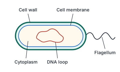
What a bacterium has
- Even the simplest bacterium has three essential parts.
- Its genetic material, usually one loop of DNA, holds the instructions that run the cell.
- A cell membrane surrounds it and controls which substances get in and out, such as letting food in and waste out.
- The cytoplasm is the jelly-like fluid filling the cell, where most of its chemical reactions happen.
- Most bacteria also have a cell wall outside the membrane, and some have a whip-like tail called a flagellum for swimming.
Organelles in plant and animal cells
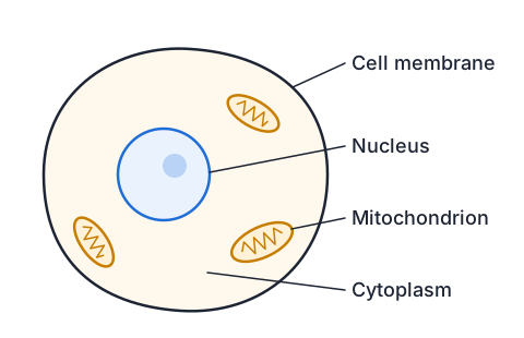
- Eukaryotic cells have the same membrane and cytoplasm as bacteria, plus organelles wrapped in their own membranes.
- The nucleus holds the DNA, which carries the instructions for how the cell is built and what it does.
- Mitochondria break down glucose using oxygen in aerobic respiration, releasing the energy the cell needs. Plant cells have them too, not only animal cells.
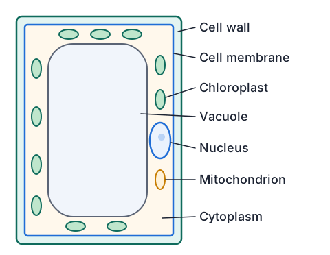
- Plant cells add three more structures: a cell wall, chloroplasts and a large permanent vacuole.
| Part | Job | Animal cell | Plant cell |
|---|---|---|---|
| Cell membrane | controls what enters and leaves | Yes | Yes |
| Cytoplasm | where many chemical reactions take place | Yes | Yes |
| Nucleus | holds DNA and controls the cell | Yes | Yes |
| Mitochondria | release energy from glucose (respiration) | Yes | Yes |
| Cell wall | made of cellulose; gives strength and a fixed shape | No | Yes |
| Chloroplasts | trap light to make glucose (photosynthesis) | No | Yes, in green parts |
| Vacuole | fluid-filled store of water, sugars and salts | small, if any | one large, permanent |
Why plant cells need the extras
- A plant has no skeleton, so it is held up by its cells: the stiff cellulose wall stops each cell bursting and gives it a fixed, boxy shape.
- A large vacuole full of water presses outwards against the wall, keeping the cell firm. When a plant runs short of water, its vacuoles shrink and it wilts.
- Chloroplasts contain the green pigment chlorophyll, which absorbs light energy to make glucose from carbon dioxide and water.
- Root cells underground get no light, so they have no chloroplasts, even though they are still plant cells.
- Animal cells have no wall, so their shapes vary and can change, from round red blood cells to long nerve cells.
Measuring cells from an image
- A microscope image shows a cell much bigger than it really is, and the magnification tells you how many times bigger.
- Magnification = image size ÷ actual size, so actual size = image size ÷ magnification.
- Both sizes must be in the same unit before you divide; convert mm to μm by multiplying by 1,000.
- A school light microscope magnifies up to about 400 to 1,000 times, while an electron microscope can magnify more than 1,000,000 times and show the inside of mitochondria.
| Cell | Typical real size | Seen with |
|---|---|---|
| Bacterium such as E. coli | about 2 μm long | light microscope at highest power (just), electron microscope for detail |
| Red blood cell | about 7 to 8 μm across | light microscope |
| Human cheek cell | about 50 to 60 μm across | light microscope |
| Onion skin cell | a few hundred μm long | light microscope, even at low power |
How big is that cheek cell?
- A photo of a cheek cell taken at ×400 shows the cell 24 mm across.
- Find the real width in micrometres.
- actual size = image size ÷ magnificationrearranged from magnification = image ÷ actual
- 24 mm = 24,000 μm1 mm = 1,000 μm
- 24,000 μm ÷ 400 = 60 μmdivide by the magnification
- The cheek cell is 60 μm across
- Typical cheek cells are 50 to 60 μmthe answer is sensible
What magnification shows a bacterium 10 mm long?
- A bacterium 2 μm long appears 10 mm long in a textbook image.
- Find the magnification, then decide which microscope took it.
- 10 mm = 10,000 μmsame unit as the actual size
- magnification = 10,000 μm ÷ 2 μmimage ÷ actual
- = ×5,000magnification has no unit
- The image is ×5,000, beyond a light microscope, so it came from an electron microscope
How microscopes revealed the cell
- In 1665 Robert Hooke looked at thin slices of cork through his microscope and named the tiny boxes he saw cells.
- In the 1670s Antonie van Leeuwenhoek, using single lenses he ground himself, became the first person to see bacteria and other single-celled life.
- By 1839 Schleiden and Schwann, having studied many plants and animals under better microscopes, proposed that all living things are made of cells.
- Light microscopes cannot show anything smaller than about 0.2 μm clearly, so the details inside mitochondria and chloroplasts stayed hidden.
- The electron microscope, built in the 1930s, uses a beam of electrons instead of light and finally showed the folded membranes inside organelles, changing how scientists understood what each part does.
Telling cells apart from images
- Three microscope images are labelled A, B and C. A shows boxy cells with thick edges and green dots. B shows a cell with no nucleus, about 2 μm long. C shows a round cell with a dark nucleus and no wall.
- Identify each from the structures you can see.
- A: cell wall + chloroplastsonly plant cells have both
- A is a plant cell, such as a leaf cell
- B: no nucleus, 2 μm longloose DNA and bacterial size mean prokaryote
- B is a bacterium
- C: nucleus, no wall, no chloroplastseukaryote without plant features
- C is an animal cell, such as a cheek cell
Common mistakes
- Animal cells have a cell wall too, just a thinner one.Animal cells have only a cell membrane; the stiff cellulose wall is found in plant cells, which is why animal cells can change shape.
- Bacteria have no nucleus, so they have no DNA.Bacteria do have DNA, usually one loop loose in the cytoplasm; having no nucleus only means it has no membrane around it.
- Actual size = image size × magnification.Divide instead: actual size = image size ÷ magnification, because the real cell is smaller than its image.
- 24 mm ÷ 400 = 0.06, so the cell is 0.06 μm across.The answer is in the unit you divided, so it is 0.06 mm, which is 60 μm; convert to μm before dividing to avoid this.
Make a lettuce leaf go floppy
You need: 2 similar lettuce leaves, 2 bowls, cold tap water, salt, a tablespoon
- Fill both bowls with cold tap water and stir 3 heaped tablespoons of salt into one of them until most of it dissolves.
- Bend each lettuce leaf gently to feel how crisp it is, then push one under the water in each bowl.
- Leave them for 20 to 30 minutes.
- Lift both leaves out and bend each one gently again.
- Look for the difference: the leaf from plain water is still crisp, but the one from salty water is soft and floppy, because water left its cells, the vacuoles shrank and stopped pressing out against the cell walls.
Safety: Salty water stings in cuts and eyes, so keep wet hands away from your face and rinse your hands when you finish.
Your turn
- 1. A student says plant cells have chloroplasts instead of mitochondria. Explain what is wrong.
- 2. A cell has DNA, a membrane, cytoplasm and a cell wall, but no nucleus. Is it prokaryotic or eukaryotic, and what kind of organism is it likely to be?
- 3. An onion cell is 300 μm long. How long will it look in an image at ×100, in mm?
- 4. Why does a lettuce left without water go floppy? Use the words vacuole and cell wall.
- 5. Why did scientists need the electron microscope to understand how mitochondria work?
Show answer
1. Plant cells have both. Mitochondria release energy from glucose in every plant cell, and chloroplasts make that glucose in the green parts. 2. Prokaryotic, because the DNA is not inside a nucleus; it is most likely a bacterium. 3. 300 μm × 100 = 30,000 μm = 30 mm. 4. Without water the large vacuoles shrink and stop pushing out against the cell walls, so the cells go soft and the leaves droop. 5. Mitochondria are only about 1 μm across and their inner folds are far smaller than the 0.2 μm limit of a light microscope; only an electron microscope magnifies and resolves enough to show them.
What to take away
- Organelles are structures inside a cell, each with its own job.
- Prokaryotic cells, such as bacteria, have no nucleus; eukaryotic cells keep their DNA inside one.
- Every cell has genetic material, a membrane that controls entry and exit, and cytoplasm where reactions happen.
- Eukaryotic cells add a nucleus and mitochondria; plant cells also have a cellulose wall, chloroplasts and a large vacuole.
- Actual size = image size ÷ magnification, with both sizes in the same unit.
- Each better microscope revealed more of the cell, from Hooke's cork boxes to the membranes inside mitochondria.
How is Year 9 maths going?
See the Year 9 maths guide