Spectroscopic data: identifying organic compounds
What this lesson covers
- Mass spectra: the molecular ion gives the molar mass, fragment peaks show pieces of the molecule, and M + 2 peaks reveal chlorine or bromine.
- Infrared spectra: bonds absorb at characteristic wavenumbers, so O–H, N–H, C=O, C=C and C–O show which functional groups are present.
- Carbon-13 NMR: the number of peaks is the number of carbon environments, and the chemical shift shows what each carbon is bonded to.
- Combining the three sets of data to decide between structural isomers and justify one structure.
Three techniques, three questions
- Each technique answers a different question about an unknown organic compound, and none of them is enough on its own.
- For this standard the compounds come from the organic families you have met: hydrocarbons (alkanes and alkenes), alcohols, haloalkanes and amines, plus the C=O families: aldehydes, ketones, carboxylic acids, esters, amides and acid chlorides.
- You are given the spectra and data tables in the assessment; the skill is reading them and linking each feature to part of the structure.
| Technique | What it measures | What it tells you |
|---|---|---|
| Mass spectrometry (MS) | Mass to charge ratio (m/z) of ions | Molar mass, and pieces of the molecule |
| Infrared (IR) | Which wavenumbers of infrared are absorbed | Bonds present, so functional groups |
| Carbon-13 NMR | Chemical shift of each kind of carbon | Number of carbon environments and their neighbours |
Mass spectrometry: the molecular ion and fragments
- The sample is vaporised and hit by high-energy electrons, which knock an electron off each molecule to make a positive molecular ion, M⁺.
- Ions carry a charge of 1+, so the peak furthest to the right (apart from small isotope peaks) is at the molar mass of the compound.
- Many molecular ions break apart; only the charged fragments are detected, and the gap between M⁺ and a fragment tells you what was lost (a gap of 15 means a CH₃ was lost).
- The tallest peak is the base peak, set to 100%: it is the most stable fragment, not necessarily the molecular ion.
- Chlorine is ³⁵Cl and ³⁷Cl in a 3 : 1 ratio, so a chloroalkane shows M and M + 2 peaks of heights 3 : 1; bromine (⁷⁹Br and ⁸¹Br) gives two peaks of equal height.
| m/z | Likely fragment ion |
|---|---|
| 15 | CH₃⁺ |
| 29 | C₂H₅⁺ or CHO⁺ |
| 31 | CH₂OH⁺ (from many alcohols) |
| 43 | C₃H₇⁺ or CH₃CO⁺ |
| 45 | COOH⁺ |
| 57 | C₄H₉⁺ or C₂H₅CO⁺ |
Example: the mass spectrum of butanone
- Butanone is CH₃COCH₂CH₃, molecular formula C₄H₈O. Its mass spectrum has peaks at m/z 72, 57, 43 and 29, with the base peak at 43.
- Explain each peak.
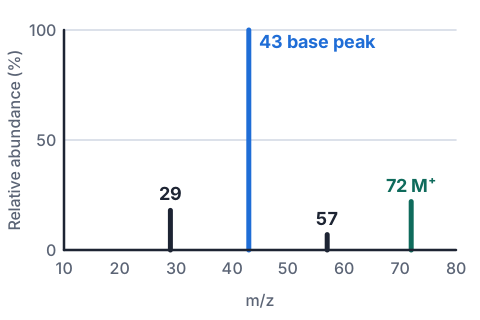
- M = 4(12.0) + 8(1.0) + 16.0 = 72.0 g mol⁻¹, so m/z 72 is M⁺, [CH₃COCH₂CH₃]⁺
- 72 − 57 = 15: loss of CH₃, leaving CH₃CH₂CO⁺
- 72 − 43 = 29: loss of C₂H₅, leaving CH₃CO⁺the base peak: the C=O stabilises this ion
- m/z 29 is C₂H₅⁺, the other half of the same break
- breaking either C–C bond next to the C=O is typical of ketones
Infrared spectra and functional groups
- Covalent bonds stretch and bend, and each kind of bond absorbs infrared at its own range of wavenumbers (cm⁻¹); the spectrum shows transmittance, so an absorption is a dip.
- The scale runs from 4000 cm⁻¹ on the left to about 500 cm⁻¹ on the right, and the useful dips for this standard are above 1500 cm⁻¹, plus C–O.
- Below 1500 cm⁻¹ is the fingerprint region: too many overlapping dips to assign, but unique to each compound, so it can match an unknown to a known spectrum.
- Hydrogen bonding broadens O–H dips: an alcohol's O–H is a broad rounded dip, and a carboxylic acid's is very broad, running into the C–H dips.
| Bond | Wavenumber (cm⁻¹) | Look of the dip | Found in |
|---|---|---|---|
| O–H | 3200 to 3550 | Broad, rounded | Alcohols |
| O–H | 2500 to 3300 | Very broad | Carboxylic acids |
| N–H | 3300 to 3500 | Medium, sharper than O–H | Amines, amides |
| C–H | 2850 to 3000 | Sharp | Almost every organic compound |
| C=O | 1650 to 1750 | Strong, sharp | Aldehydes, ketones, acids, esters, amides, acid chlorides |
| C=C | 1620 to 1680 | Medium to weak | Alkenes |
| C–O | 1000 to 1300 | Strong | Alcohols, acids, esters |
Example: acid, alcohol or aldehyde?
- A bottle could hold propan-1-ol, propanal or propanoic acid. Its IR spectrum has a very broad dip from 2500 to 3300 cm⁻¹, a strong sharp dip at 1710 cm⁻¹ and a dip at 1240 cm⁻¹.
- Which compound is it?
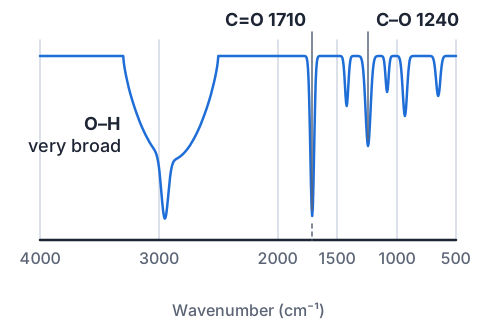
- 1710 cm⁻¹ strong and sharp: C=O presentrules out propan-1-ol, which has no C=O
- very broad 2500 to 3300 cm⁻¹: O–H of a carboxylic acidrules out propanal, which has C=O but no O–H
- 1240 cm⁻¹: C–O, as in COOH
- the compound is propanoic acid, CH₃CH₂COOH
Try it: which bond made that dip?
Carbon-13 NMR: counting carbon environments
- In a magnetic field, each ¹³C nucleus absorbs radio waves at a frequency set by the atoms around it, reported as a chemical shift δ in ppm from 0 to about 220.
- Carbons in identical surroundings give one peak, so the number of peaks is the number of carbon environments, not the number of carbons.
- Look for symmetry: in propan-2-ol, CH₃CH(OH)CH₃, the two CH₃ carbons are equivalent, so three carbons give only two peaks.
- The shift shows the neighbours: a carbon bonded to an electronegative atom such as O or Cl is pulled to a higher δ, and a C=O carbon sits highest of all.
- Peak heights in a ¹³C spectrum do not reliably show how many carbons are in each environment, so use the number and the positions of peaks, not their heights.
| Kind of carbon | Chemical shift δ (ppm) |
|---|---|
| C–C in an alkyl chain (CH₃, CH₂, CH) | 0 to 50 |
| C–Cl or C–Br | 30 to 70 |
| C–N | 30 to 65 |
| C–O (alcohols, ethers, esters) | 50 to 90 |
| C=C | 110 to 150 |
| C=O in acids, esters, amides, acid chlorides | 160 to 185 |
| C=O in aldehydes and ketones | 190 to 220 |
Example: telling the butanol isomers apart
- Four alcohols share the formula C₄H₁₀O, so their mass spectra all show M⁺ at m/z 74 and their IR spectra all show a broad O–H dip.
- Predict the number of peaks in each ¹³C NMR spectrum.
| Alcohol | Structure | Carbon environments | Peaks |
|---|---|---|---|
| Butan-1-ol | CH₃CH₂CH₂CH₂OH | every carbon different | 4 |
| Butan-2-ol | CH₃CH(OH)CH₂CH₃ | every carbon different | 4 |
| 2-methylpropan-1-ol | (CH₃)₂CHCH₂OH | two CH₃ the same | 3 |
| 2-methylpropan-2-ol | (CH₃)₃COH | three CH₃ the same | 2 |
- 2 peaks: only 2-methylpropan-2-ol
- 3 peaks: only 2-methylpropan-1-ol
- 4 peaks: butan-1-ol or butan-2-ol, so look at the C–O carbon and the fragmentsbutan-1-ol gives a strong m/z 31 (CH₂OH⁺); butan-2-ol gives m/z 45 (CH₃CHOH⁺)
- the peak count alone separates two of the four; MS fragments separate the other two
Try it: count the carbon environments
Combining the data
- Start with the mass spectrum: M⁺ gives the molar mass, and with the elements present it fixes the molecular formula.
- Use the IR spectrum to name the functional group, and say which groups are ruled out by missing dips.
- Draw every structural isomer with that formula and group, then use the ¹³C NMR peak count and shifts to cut the list to one.
- Check the answer against all the data: the fragments in the mass spectrum must be pieces your structure can actually make, and every NMR peak must belong to a carbon in it.
Example: naming compound X
- Compound X contains carbon, hydrogen and oxygen. MS: M⁺ at m/z 88, with peaks at 73, 45 and 43. IR: very broad dip from 2500 to 3300 cm⁻¹ and a strong dip at 1710 cm⁻¹. ¹³C NMR: three peaks, at 184, 34 and 19 ppm.
- Identify X and justify it from all three spectra.
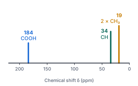
- IR: very broad O–H and C=O at 1710 cm⁻¹, so X is a carboxylic acid, R–COOH
- M = 88: COOH is 45, so R is 43, which is C₃H₇; X is C₄H₈O₂4(12.0) + 8(1.0) + 2(16.0) = 88.0
- isomers: butanoic acid CH₃CH₂CH₂COOH (4 environments) or 2-methylpropanoic acid (CH₃)₂CHCOOH (3 environments)
- NMR shows 3 peaks: 184 ppm COOH, 34 ppm CH, 19 ppm two equivalent CH₃rules out butanoic acid
- MS: 88 − 15 = 73 (loss of CH₃), 45 is COOH⁺, 43 is (CH₃)₂CH⁺
- X is 2-methylpropanoic acid, (CH₃)₂CHCOOH
How it is assessed, and what Merit and Excellence look like
- This is an internal: you are given spectra and data tables for unknown compounds, usually under test conditions, and write your analysis.
- Achieved identifies separate features: a functional group from an IR dip, the molar mass from M⁺, the number of carbon environments.
- Merit uses the data to work out the full structure; Excellence justifies that structure by linking all three spectra and explaining why the other isomers do not fit.
| Grade | What the student wrote |
|---|---|
| Achieved | The dip at 1710 cm⁻¹ shows a C=O bond, and the molecular ion at 88 means the molar mass is 88 g mol⁻¹. |
| Merit | The very broad O–H and the C=O show a carboxylic acid. With M = 88 the formula is C₄H₈O₂, and three ¹³C peaks fit 2-methylpropanoic acid, (CH₃)₂CHCOOH. |
| Excellence | IR gives a carboxylic acid, since the O–H dip is very broad and C=O is at 1710 cm⁻¹. M⁺ at 88 makes the formula C₄H₈O₂, so X is butanoic acid or 2-methylpropanoic acid. Butanoic acid has four carbon environments, but X shows three: 184 ppm (COOH), 34 ppm (CH) and 19 ppm, one peak for two equivalent CH₃ groups. The fragments agree: 73 is M⁺ minus CH₃ and 43 is (CH₃)₂CH⁺. X is 2-methylpropanoic acid. |
Common mistakes
- The tallest peak in the mass spectrum is the molecular ion, so M = 43.The tallest peak is the base peak, the most common fragment; the molecular ion is the last big peak on the right.
- Propan-2-ol has three carbons, so its ¹³C NMR spectrum has three peaks.The two CH₃ groups are equivalent, so there are two carbon environments and two peaks.
- There is a dip at 3000 cm⁻¹, so the compound has an O–H group.C–H dips sit at 2850 to 3000 cm⁻¹ in almost every compound; an alcohol O–H is a broad dip at 3200 to 3550 cm⁻¹.
- A C=O dip at 1715 cm⁻¹ means it is a ketone.Aldehydes, acids, esters and amides all have C=O too; check for O–H or N–H in IR and for the C=O shift in the ¹³C spectrum.
Count carbon environments with models
You need: a stick of Blu Tack or playdough, a box of toothpicks, two colours of felt pen, paper
- Roll black-marked balls for carbon and plain smaller balls for hydrogen, and join them with toothpicks into pentane, CH₃CH₂CH₂CH₂CH₃.
- Turn the model end to end and mark carbons that look the same from their own position with the same colour; count the colours to predict the ¹³C peaks.
- Rebuild the five carbons as 2-methylbutane and then as 2,2-dimethylpropane, and count again.
- Write the three formulas (all C₅H₁₂, all M⁺ at 72) beside your peak counts.
- Look for: pentane gives 3 peaks, 2-methylbutane 4 and 2,2-dimethylpropane 2, so NMR tells apart isomers that MS and IR cannot.
Safety: Toothpick points can pierce skin or an eye, so push them in pointing away from you and keep them away from small children.
Your turn
A C₃H₈O alcohol has a broad IR dip at 3350 cm⁻¹ and two peaks in its ¹³C NMR spectrum. Name it.
How many peaks does pentan-3-one, CH₃CH₂COCH₂CH₃, give in its ¹³C NMR spectrum?
A haloalkane has molecular ion peaks at m/z 78 and 80 in a 3 : 1 ratio. Which halogen is present, and what is the molecular formula?
An ester C₄H₈O₂ has its base peak at m/z 43, a CH₃CO⁺ ion. Is it ethyl ethanoate or methyl propanoate?
Which IR evidence shows a sample is propanal and not propan-1-ol?
Show answer
1. Two peaks means two carbon environments, so it is propan-2-ol, CH₃CH(OH)CH₃ (propan-1-ol has three). 2. The molecule is symmetrical about the C=O: the two CH₃ are equivalent and the two CH₂ are equivalent, so 3 peaks (CH₃, CH₂ and C=O). 3. Two peaks 2 apart in a 3 : 1 ratio show chlorine; 78 − 35 = 43 = C₃H₇, so C₃H₇Cl. 4. Ethyl ethanoate, CH₃COOCH₂CH₃, which breaks to CH₃CO⁺ (m/z 43); methyl propanoate would give CH₃CH₂CO⁺ at m/z 57. 5. A strong sharp C=O dip near 1730 cm⁻¹ and no broad O–H dip at 3200 to 3550 cm⁻¹.
Recap
- MS: M⁺ gives the molar mass, gaps to fragments show what broke off, and M + 2 peaks flag Cl (3 : 1) or Br (1 : 1).
- IR: O–H broad (very broad in acids), N–H, C=O strong near 1700 cm⁻¹, C=C, and C–O; the fingerprint region matches but is not assigned.
- ¹³C NMR: one peak per carbon environment; the shift rises with nearby O, N or halogen and is highest for C=O.
- Combine: formula from MS, group from IR, isomer from NMR, then check every peak against your structure.
| Data | Key question |
|---|---|
| Molecular ion | What is the molar mass? |
| Fragments | Which pieces break off? |
| IR dips | Which functional groups are there, and which are missing? |
| ¹³C peak count | How many carbon environments? |
| ¹³C shifts | What is each carbon bonded to? |
Report a mistake
How is NCEA Level 3 maths going?
See the NCEA Level 3 maths guide